


Anyone who wants to know how far their own hair loss has progressed sooner or later comes across the Norwood scale. It sorts male pattern hair loss into a picture-based system of seven stages plus the type A variants. O’Tar Norwood published this version in 1975 in the Southern Medical Journal after examining 1,000 men, and it is still the benchmark in everyday clinical practice. Here you can see every stage illustrated, how to place yourself on the scale and what your stage means for treatment.
The key points
- ✓Stage 1 is the youthful hairline, stage 7 leaves only a narrow rim of hair above the ears and at the nape
- ✓Two zones decide the stage: the front hairline and the crown
- ✓The scale describes a visible pattern. It is not a diagnosis and not a prognosis
- ✓Women are assessed with the Ludwig scale and its three grades, because female hair loss follows a different pattern
The table of contents shows you what is covered where. After that we go into detail, stage by stage.
Summary
- What is the Norwood scale?
- How to work out your Norwood stage yourself
- Norwood 1: the unchanged youthful hairline
- Norwood 2: slight recession at both temples
- Norwood 3 and Norwood 3 vertex: the first stage that is relevant for treatment
- Norwood 4: frontal zone and crown, separated by a narrow band of hair
- Norwood 5: the bridge of hair between the zones grows thin
- Norwood 6: frontal zone and crown are connected
- Norwood 7: only the rim of hair remains
- The type A variant of the Norwood scale: when the hairline moves back evenly
- The Norwood scale at a glance: all stages in one table
- What follows from my Norwood stage? Treatment by stage
- The three biggest misconceptions about the Norwood scale
- Limits of the Norwood scale: what it cannot do
- Ludwig scale: the classification for women
- Having your Norwood stage determined by a doctor
- When the Norwood scale does not fit: warning signs and differentiation
- Frequently asked questions about the Norwood scale
- Sources
What is the Norwood scale?
The Norwood scale is a picture-based system that doctors use to sort male pattern hair loss into seven clearly described stages. It gives doctors, patients, research and transplant planning a shared language for a visible pattern.
What the scale shows is the result of a hormonal process. In androgenetic alopecia, dihydrotestosterone (DHT) binds to the androgen receptor of genetically sensitive follicles at the forehead, temples and crown. These follicles miniaturise: with every cycle the hair becomes thinner, shorter and lighter, until it stops growing altogether.
At the back and sides of the head the follicles remain largely DHT-resistant, which is why hair stays there and why this is precisely the area that serves as the donor zone (British Association of Dermatologists, patient information on male pattern hair loss). It is only because the loss always follows the same zones that it can be divided into stages at all.
Two directions of progression are typical: from the front across the temples as frontotemporal recession, in other words receding temples, and outwards from the crown as a bald spot at the crown. In the higher Norwood stages the two fronts meet.
What the Norwood scale does not do: it identifies no cause, no pace and no point in time. It says nothing about hair density, hair calibre or the degree of miniaturisation. It is a tool for communication, not a diagnosis.
Norwood scale or Hamilton-Norwood scale? One classification, two names
Both terms describe the same classification. James B. Hamilton published the first systematic classification of the male hair loss pattern in 1951 in the Annals of the New York Academy of Sciences. O’Tar Norwood, a dermatologist and hair transplant surgeon, revised it in 1975 in the Southern Medical Journal on the basis of 1,000 men he had examined.
Norwood added the separate vertex stage and the type A variants. You will find the scale called the “Norwood scale”, the “Hamilton-Norwood scale” or the “Norwood-Hamilton scale”; medically these are one and the same.
How to work out your Norwood stage yourself
For a reliable self-assessment on the Norwood scale you need three photos taken in the same light: the forehead from the front, the head from above and a profile shot. Anyone who only estimates their stage in the mirror almost always underestimates the crown, simply because they cannot see it.
The real trick lies in the way the scale is constructed. Norwood defines the stages through two independent zones, the front hairline and the crown. Assess both separately and then combine them, and you will land on the right stage far more reliably than by comparing rows of pictures.
| Assessing the hairline | Crown: full | Crown: visibly thinning | Crown: bald |
|---|---|---|---|
| Straight, no recession | Stage 1 | Thinning without recession: not cleanly captured by Norwood, have it checked by a doctor | unusual, have it checked by a doctor |
| Slight recession at both temples | Stage 2 | Stage 2 to 3 vertex | unusual, have it checked by a doctor |
| Deep, wedge-shaped M recession | Stage 3 | Stage 3 vertex | Stage 4 |
| Frontal zone largely bald, band of hair still standing | check for a type A pattern | Stage 4 | Stage 4 to 5 |
| Frontal zone bald, band of hair thin or gone | check for a type A pattern | Stage 5 | Stage 6 to 7 |
Two notes on the matrix: if you see a line that has moved back evenly without an M shape, the section on the type A variant is the one to read. And remember that the matrix classifies a visible pattern: it is not a diagnosis and it does not capture miniaturisation.
Photo protocol: how a snapshot becomes a documented progression
- 1.Photograph three angles: forehead from the front, head from above (crown), profile.
- 2.Always dry hair. Wet hair makes any hairline look thinner than it is.
- 3.Daylight from the side, no ceiling spotlight directly above your head.
- 4.Push the hair back off the forehead, raise your eyebrows as far as they go and use the highest crease as your reference line.
- 5.Same distance, same angle, same camera every time you repeat it.
- 6.Put the date in the file name and repeat every 6 months.
One set of photos records a state. Two sets, 6 to 12 months apart, show a progression, which is exactly what a single Norwood picture cannot do.
The most common error in self-assessment: a single photo shows a state, not a progression. Only comparing two sets of photos taken 6 to 12 months apart shows whether there is active hair loss at all. That is precisely what decides whether you are looking at a mature hairline or an early stage 2.
The second blind spot is miniaturisation. Thinner, shorter and lighter hairs in one zone are an earlier signal than a visible gap, and they appear in no Norwood picture. A scalp magnifier or a medical trichoscopy makes them visible.
And the honest limitation: in a study in which 7 dermatologists and 16 trainee doctors classified 43 photographs, agreement was insufficient (ICC 0.63 to 0.68, International Journal of Trichology 2009). Even the same assessors reached different results 3 months later. So your self-assessment is a useful starting point, not a measurement.
Norwood 1: the unchanged youthful hairline
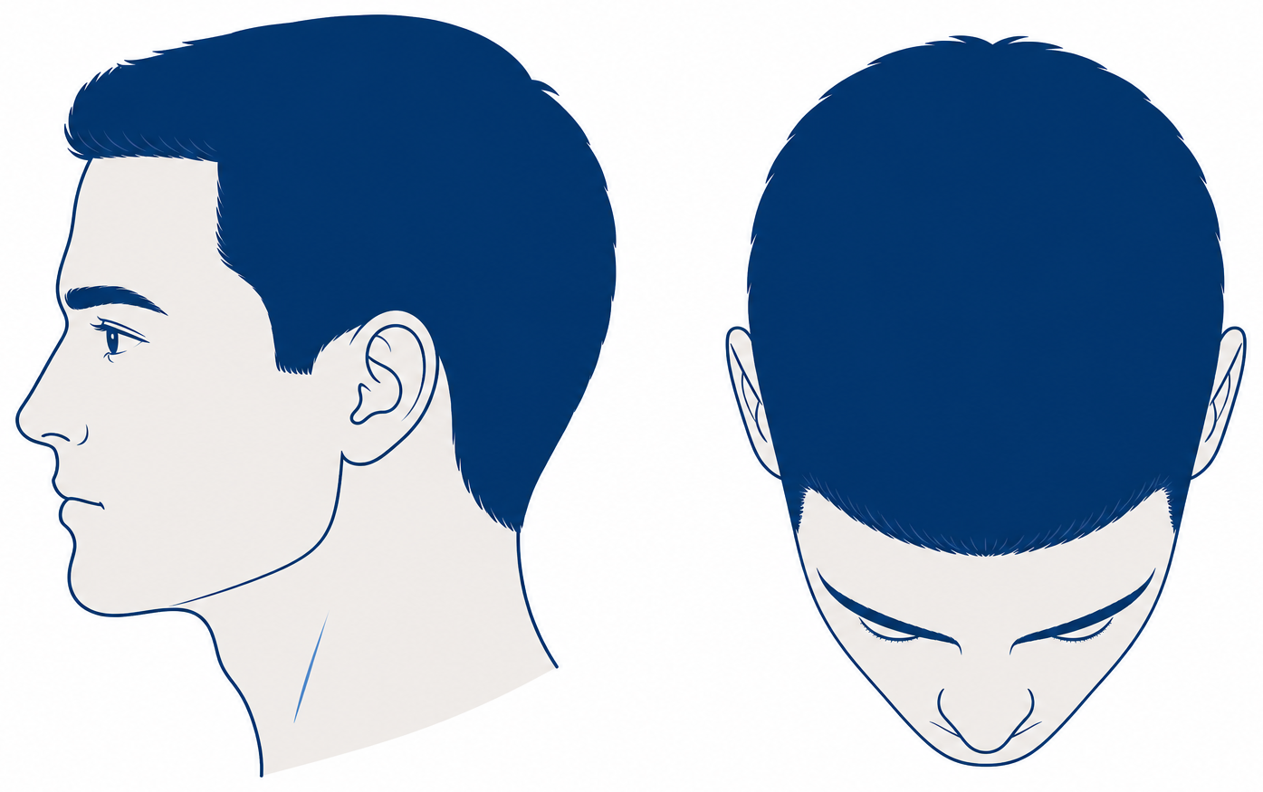
Norwood 1 describes a hairline with no visible recession, the way it runs in youth. It is the reference stage of the scale and not a stage of hair loss.
In the mirror you see a straight or minimally curved line across the forehead, no wedge-shaped gaps at the temples, and on the top-down photo a fully covered crown. In the Japanese cohort study of 5,372 men the median age at stage I was 26 years (95 per cent confidence interval 25 to 27). That is an observation in this cohort and not an expectation for you.
Graft requirement: none. At stage 1 there is no indication for a hair transplant. The sensible next step is unspectacular but valuable: take reference photos following the protocol above. If you document your starting point at 20, you can later show what has changed instead of guessing.
Norwood 2: slight recession at both temples
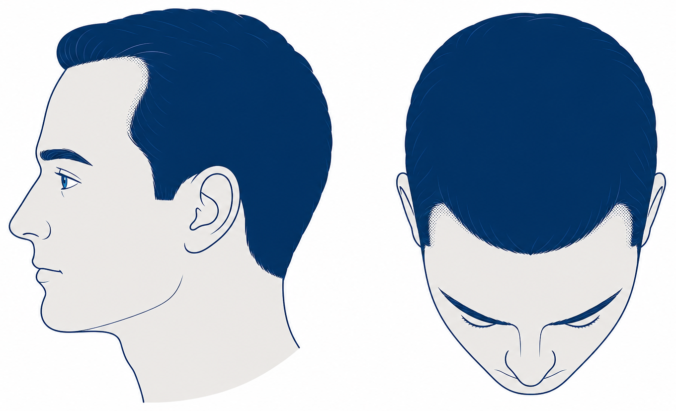
Norwood 2 means a slight, usually symmetrical recession of the hairline at both temples. The hairline has moved back by a few millimetres to around a centimetre, the forehead looks a little higher, and the top of the head is fully covered.
What you see is the first hint of the M shape. The remaining line still runs without a break, and there is no visible thinning at the crown. This is exactly where the key question of this stage comes up.
Norwood 2 or just a mature hairline?
A mature hairline moves back once, by roughly 1 to 1.5 centimetres, during puberty and the early twenties and then stays stable, with no miniaturisation. Norwood 2 in the sense of active hair loss keeps progressing and shows thinner, shorter, lighter hairs along the edge. This distinction is a matter of broad clinical consensus, not the result of a single study.
On a single photo the two cannot be told apart. Only a comparison over time answers the question. Norwood 2 at the age of 20 is possible and no emergency in itself, but it is a good reason to document the progression now and have it assessed by a dermatologist: hair loss that starts early tends on average to progress further.
If surgery is considered at this stage at all, it involves a small area in the region of 1,000 to 1,500 grafts. That is a rough guide, not a promise: it depends on the individual donor area at the back of the head, it shifts with hair calibre, because fine hair covers less area per graft, and it is set solely by the medical examination.
So the sensible next step is usually not a procedure. At stage 2, a proper work-up and, where progression is documented, stabilisation with medication take priority. If grafts go into an actively progressing area, the transplanted hairs stay while the native hair around them keeps falling out. Surgeons describe this result as the island effect. Details on the frontal zone can be found in the article on receding temples.
Norwood 3 and Norwood 3 vertex: the first stage that is relevant for treatment
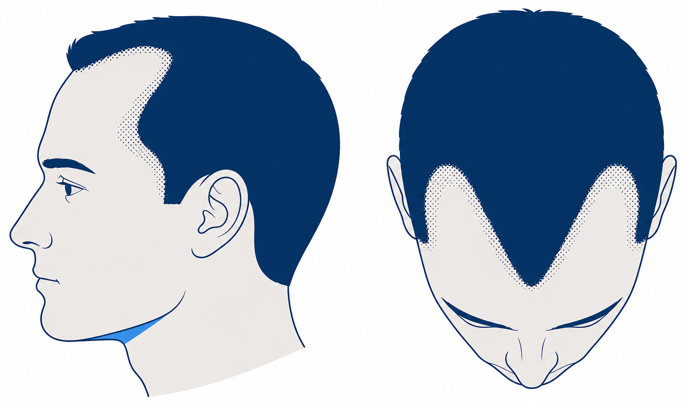
By Norwood’s own definition, Norwood 3 is the first stage that counts as hair loss relevant for treatment. The temples have receded clearly and deeply in a wedge shape, the M shape is unmistakable, and the remaining line in the middle is in places only narrow.
Norwood 3 vertex is clearly separate from this: the hairline has receded as it does at stage 3 and, in addition, the crown on top of the head is thinning, while a band of hair still stands between the two zones. On the top-down photo the scalp shows through there. That is two areas to address instead of one.
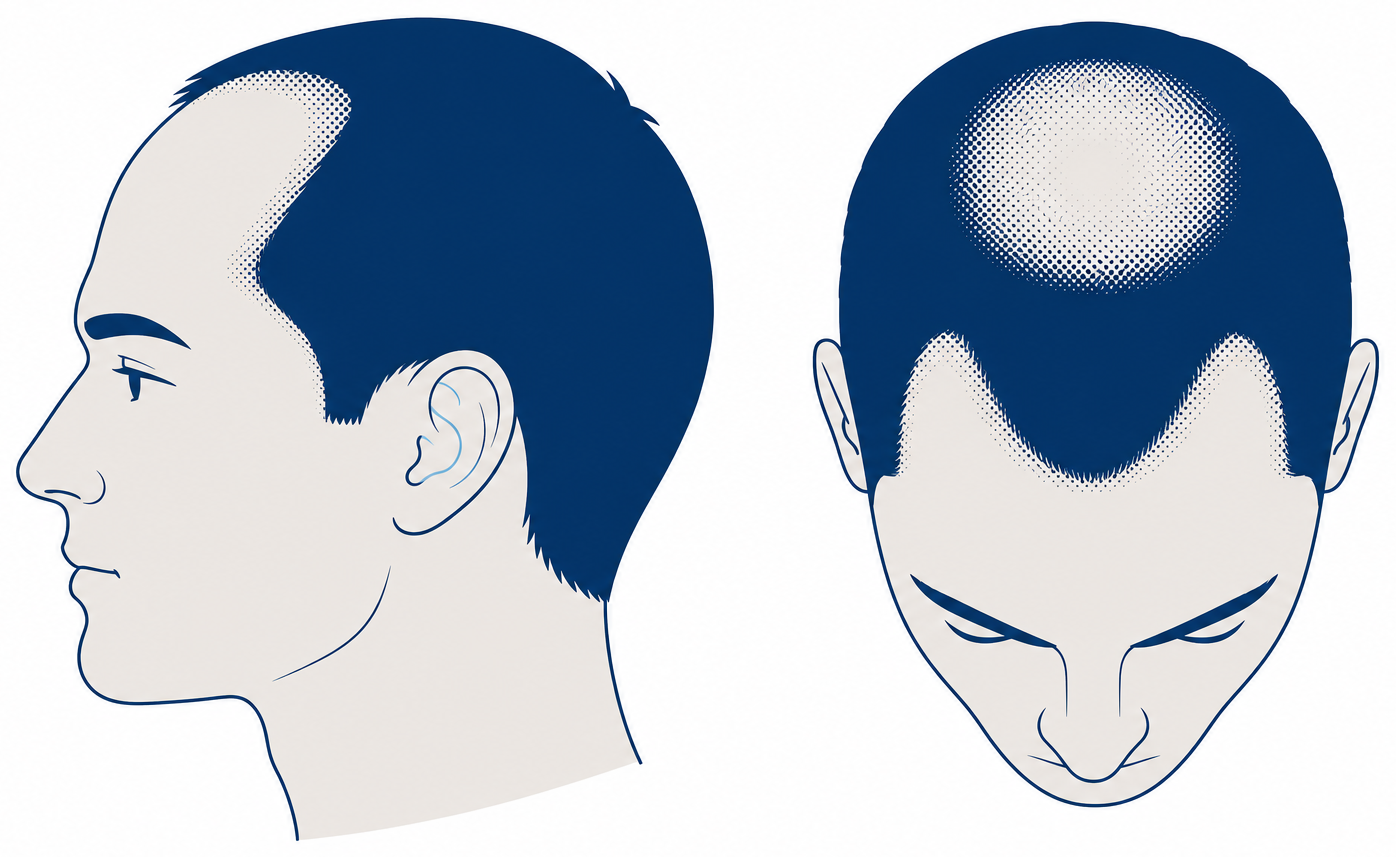
Norwood 3 is one of the most common classifications in middle adulthood in many cohorts. A review in the Indian Journal of Dermatology, Venereology and Leprology puts the figures at around 30 per cent of Caucasian men affected in their thirties and 40 per cent in their forties. These, too, are cohort figures, not a timetable for the individual.
For the front zone the requirement is in the region of 1,500 to 3,000 grafts, and correspondingly higher for Norwood 3 vertex, because the crown is an additional area. Just as important: the donor area is limited, with fine, straight hair the requirement for the same area is considerably higher than with thick or curly hair, and the binding figure comes from the medical examination.
From here on a transplant becomes medically worth discussing, and usually makes most sense combined with stabilising the remaining hair. The real planning conflict: hairline and crown compete for the same donor hair. How the crown area can be treated is covered in the article on crown balding.
Norwood 4: frontal zone and crown, separated by a narrow band of hair
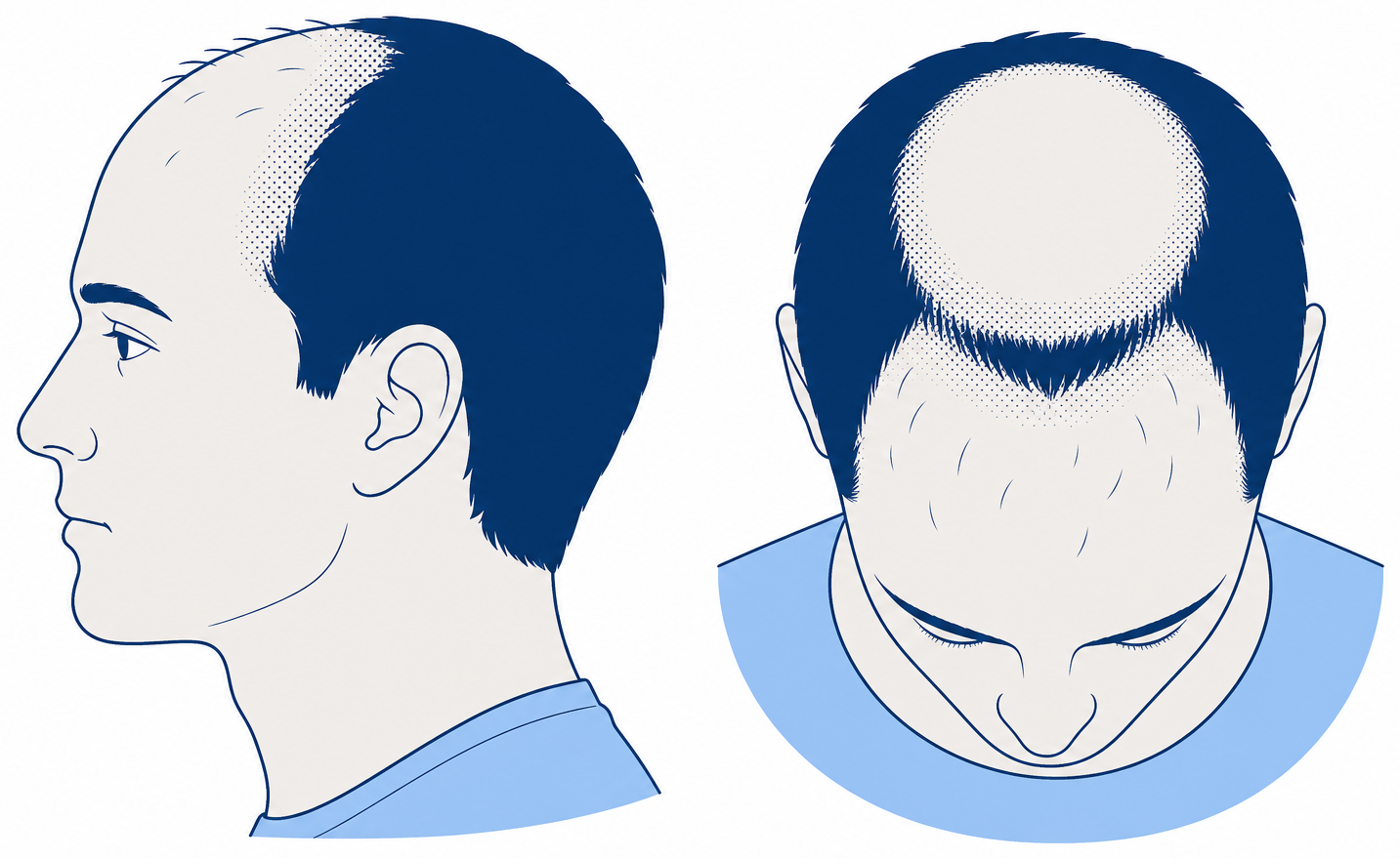
Norwood 4 means that the hairline has receded strongly and the crown is clearly thinning or bald. Between the bald frontal zone and the bald crown only a narrow band of hair remains across the top of the head.
What you see is a clearly enlarged forehead, often with only sparse single hairs in the frontal zone, a clearly visible bald area at the crown and a separating band of hair that is getting thinner. This stage is a turning point, because the bald area typically exceeds what can be covered at full youthful density.
Planning therefore becomes a question of priorities: frame and hairline first, because they define the face, the crown afterwards or at a lower density. In the Japanese cohort study of 5,372 men the median time from one stage to the next was 4.5 years, and higher stages appeared at rising median ages. Where hair loss starts early, Norwood 4 is reached considerably sooner.
The requirement is in the region of 3,000 to 4,000 grafts for a visible improvement in both zones. That range assumes a sufficiently dense, healthy donor zone, it shifts with hair calibre, and whether it is achievable in your case is decided not by the Norwood stage but by the medical examination.
Can Norwood 4 still be halted? Progression can be slowed with medication, but lost area does not grow back as a result. Preserving and restoring are two different things: medication holds on to what is there, a transplant redistributes existing hair. How much area can realistically be covered is shown in the article on possible graft numbers.
Norwood 5: the bridge of hair between the zones grows thin
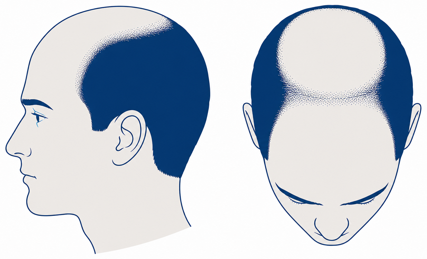
At Norwood 5 the frontal zone and the crown have grown further, and the band of hair between them has become distinctly narrower and thinner. The two bald areas are still separate, but visibly on their way towards each other.
What you see is a narrow, see-through strip of hair across the head, with the bald zones reaching further back and towards the sides. This stage is mostly observed in later adulthood, though with wide individual variation.
The requirement is in the region of 3,500 to 4,500 grafts. From Norwood 5 onwards the limiting factor is the donor area, not what you would like: clinics and specialist sources consistently give roughly 4,000 to 8,000 grafts as safely harvestable over a lifetime, often 6,000 to 7,000 as the practical upper limit. Those are benchmarks from practice, not study figures.
Full youthful density across the whole area is generally not achievable at this stage. What is realistic is a clear improvement through strategic distribution, planned individually by a doctor. Sensible goals are a hairline that suits your age and a complete visual frame, not the maximum covered area.
Norwood 6: frontal zone and crown are connected
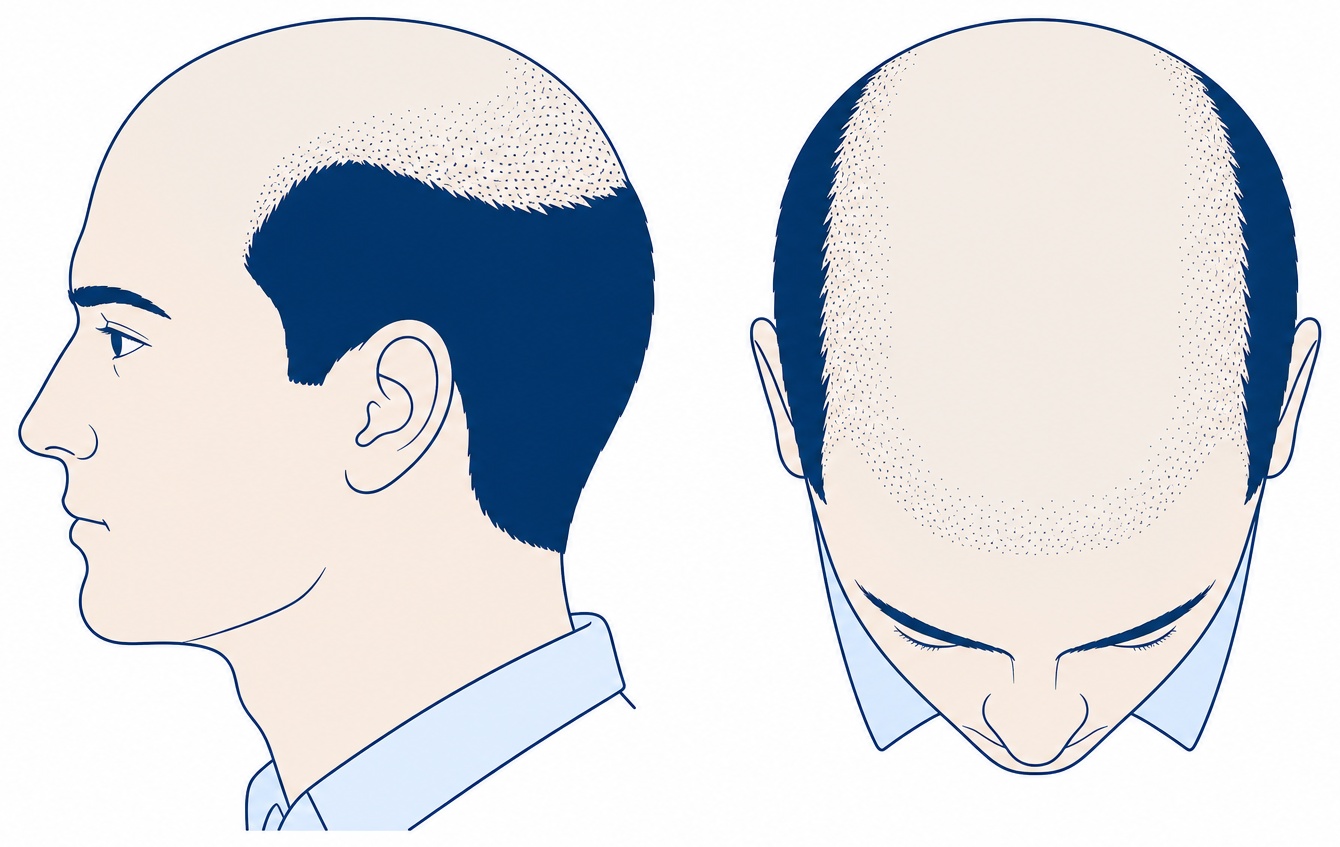
Norwood 6 is present once the separating band of hair has disappeared and the bald frontal zone merges into the bald crown. A rim of hair remains at the sides and the back of the head, and it is still relatively broad.
What you see is one continuous bald area across the top of the head. This is mostly observed at older ages: Australian cohort data, summarised in reviews, show a rise in crown or full baldness from 31 per cent among 40- to 55-year-olds to 53 per cent among 65- to 69-year-olds.
The requirement is in the region of 5,000 to 6,000 grafts and therefore exceeds the available donor reserve in many cases. Even when the donor zone is used to the maximum, this is about a visible improvement in selected zones, not complete coverage. What is possible is decided by the medical assessment of donor density and hair quality.
The sensible next step is therefore to set priorities. The hairline and frontal zone give the face its frame back, a short haircut can be planned in from the start, and alternatives should be on the table from the outset.
Norwood 7: only the rim of hair remains
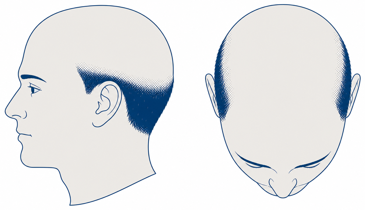
Norwood 7 is the final stage of the scale. Only a narrow rim of hair above the ears and at the nape is left, and even this rim can become thinner. The top of the head is completely bald.
In the Japanese cohort study of 5,372 men the median age at stage VII was 52 years (95 per cent confidence interval 46 to 61). With an aggressive course this stage is reached sooner; the figure describes a cohort, not an individual course.
On the graft requirement there is no figure here, but a clear statement: restoring the whole area with your own hair is generally not possible, because the donor area itself is reduced. If you still want to change something, there are options for selected sub-areas depending on the findings.
To be honest about it, the non-surgical routes belong here too: a very short cut or shaving as a deliberate decision, scalp micropigmentation (SMP) to thicken the rim visually, or a hair system. Whether any surgical option remains at all is something only the medical examination can clarify.
The type A variant of the Norwood scale: when the hairline moves back evenly
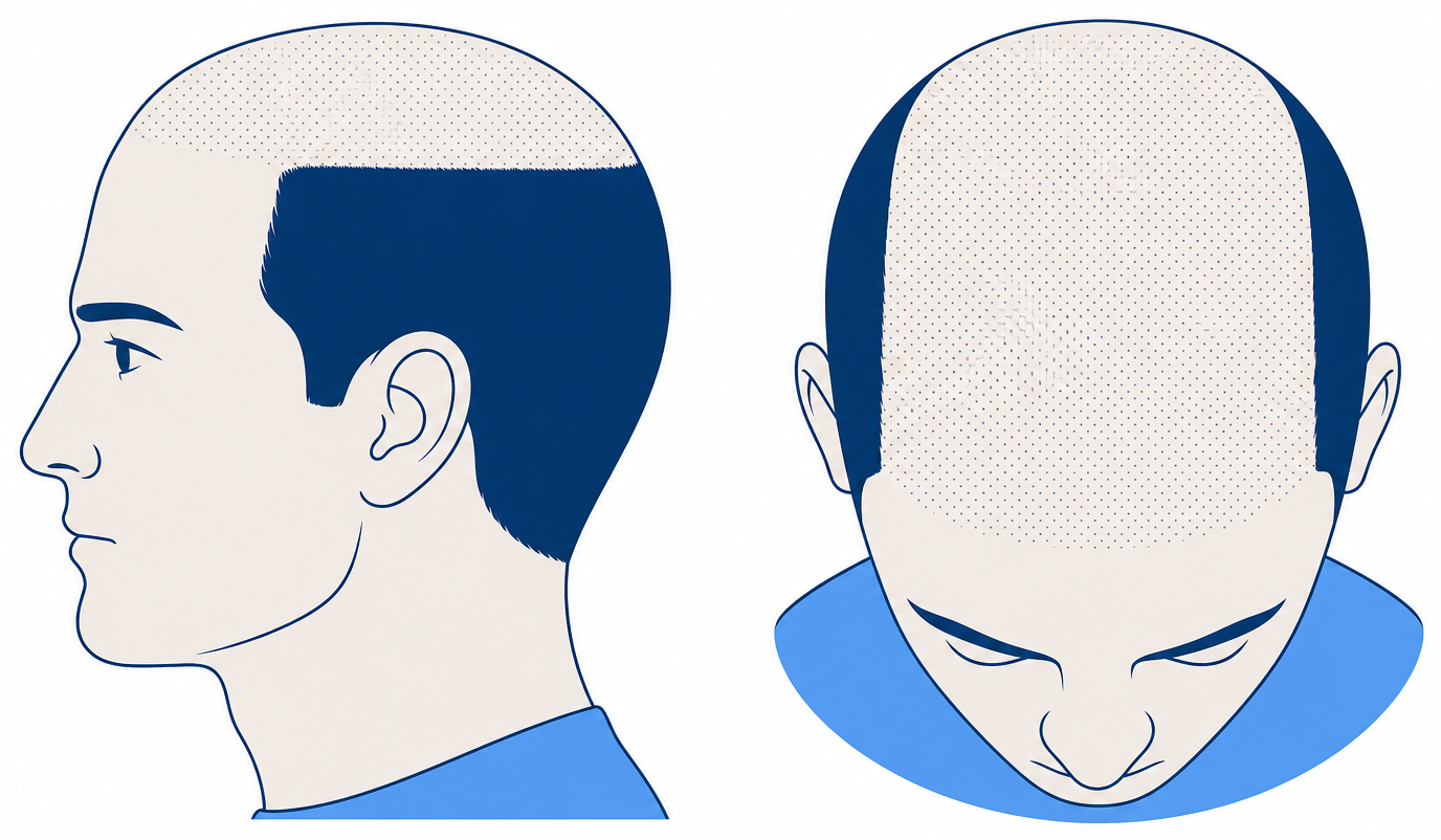
In the type A variants of the Norwood scale (IIa, IIIa, IVa, Va) the hairline moves back evenly as a whole, without the typical wedge-shaped M form. The loss runs as one front from the front of the head backwards instead of starting at two separate points.
Important and often misrepresented: the difference is not that the crown is spared. In type A no isolated, separate bald spot appears at the crown as it does in the standard pattern. In the higher type A grades the moving front does reach the mid-scalp and crown area, just coming from the front and not as a second, independent site.
Secondary sources consistently put the type A patterns at roughly 3 to 4 per cent of cases; there is no independent primary study on this. Those affected do not recognise themselves in the standard pictures and therefore often place themselves in the wrong stage. The tell-tale signs are the absence of wedge-shaped temple angles and a front line that has receded evenly and high.
For planning this means: the bald area is continuous and lies at the front, where a result shapes the face. At the same time it often reaches far back, which increases the requirement. The figures are comparable to those for the main stages, with the same limits in terms of donor area, hair calibre and individual medical assessment. Type A is not a worse pattern, just a different one.
The Norwood scale at a glance: all stages in one table
The table below summarises all seven Norwood stages plus the vertex variant: what you see, from when the stage is frequently observed in study cohorts, roughly how many grafts are in play and what the sensible next step is.
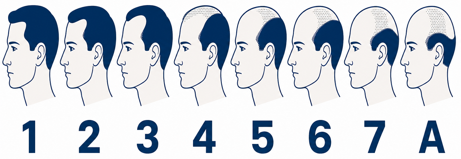
| Stage | What you see in the mirror and on the top-down photo | Frequently observed from | Approximate graft requirement | Sensible next step |
|---|---|---|---|---|
| Norwood 1 | Youthful hairline, full crown | youth, early adulthood | no requirement | take reference photos |
| Norwood 2 | Slight recession at both temples, top of the head full | often mid-twenties to early thirties | roughly 1,000 to 1,500, if at all | document the progression, get it assessed, usually no surgery yet |
| Norwood 3 | Deep, wedge-shaped M recession | middle adulthood | roughly 1,500 to 3,000 | have stage, donor area and hair calibre assessed by a doctor |
| Norwood 3 vertex | M recession plus thinning crown, band of hair between them still standing | middle adulthood | more than stage 3, because two separate zones are covered | prioritise the zones |
| Norwood 4 | Large bald frontal zone, bald crown, narrow band of hair between them | more often from the fifth decade of life | roughly 3,000 to 4,000 | plan the frame and hairline first |
| Norwood 5 | Band of hair distinctly narrower and thinner | later adulthood | roughly 3,500 to 4,500, donor area limited | clarify expectations before planning |
| Norwood 6 | Frontal zone and crown are connected, rim of hair remains | mostly older ages | roughly 5,000 to 6,000, often more than the reserve allows | define partial goals |
| Norwood 7 | Only a narrow rim of hair, partly thinned itself | old age, sooner with an aggressive course | full coverage with your own hair usually not achievable | discuss alternatives openly |
How to read the table: it offers a rough guide, not a diagnosis. The ages describe observations in study cohorts, not your own course. All graft figures are approximate and shift with hair calibre and hair texture. The donor area at the back of the head is limited and does not grow back. What is possible in your case is set solely by the medical examination.
How quickly do you progress on the Norwood scale?
The median time from one Norwood stage to the next was 4.5 years in an analysis of 5,372 Japanese men at the Tokyo Memorial Clinic. The median age rose steadily with every stage, from 26 years at stage I to 52 years at stage VII.
| Observation | Figure | Source |
|---|---|---|
| Median age at Hamilton-Norwood stage I | 26 years (95 per cent confidence interval 25 to 27) | cohort study of 5,372 Japanese men, Tokyo Memorial Clinic |
| Median age at stage VII | 52 years (95 per cent confidence interval 46 to 61) | the same cohort study, rise in median age per stage with p below 0.0001 |
| Median time from one stage to the next | 4.5 years | the same cohort study |
| Caucasian men affected by decade of life | around 30 per cent in their thirties, 40 per cent in their forties, 50 per cent in their fifties | review “Androgenetic alopecia: An update”, IJDVL |
| Affected over the course of a whole lifetime | up to 80 per cent of Caucasian men, 42 to 50 per cent of women | European S3 guideline, Kanti et al. 2018 |
These figures come from a single clinic and a single ethnic group, and the authors themselves cite clinic bias and the purely visual assessment as limitations. So they describe a pace that is frequently observed, not a rule: an aggressive course can reach the same stage two decades earlier.
What follows from my Norwood stage? Treatment by stage
The Norwood stage does not say which treatment works, but it says very precisely which expectations are realistic. Roughly speaking: the earlier the stage, the more can be preserved. The later the stage, the more it is about restoring with limited material.
Norwood 1 to 2: document the progression, have the cause medically confirmed, stabilise with medication where progression is documented. A transplant is usually premature here, because the island effect is a real risk. Norwood 3 to 4: stabilising remains the basis, and a transplant becomes medically sensible when lost area is to be brought back.
Norwood 5 to 6: planning becomes prioritising, frame and hairline first, the crown at lower density or not at all. Norwood 7: covering the whole area with your own hair is usually not achievable, and alternatives should be discussed openly. The cost question hangs on the graft number and therefore on what the examination finds, and the price overview gives a rough idea.
Medication: what finasteride and minoxidil achieve in each zone
Finasteride and minoxidil are designed for preservation and thickening, not for winning back bald areas. The European S3 guideline (Kanti et al. 2018, JEADV) lists both as therapies with a high level of evidence. Finasteride requires a prescription, and both call for medical advice about side effects.
It is worth being precise about the difference between the zones. The US approval for topical 5 per cent minoxidil rests on studies at the crown, and the labelling states that efficacy at the frontal hairline is not established (FDA label). A 104-week study by Kanti et al. (2016, JEADV) did, however, find a significant increase in non-vellus hairs in both zones with minoxidil foam.
For your planning that means: at the crown the evidence base is historically broader, at the front line it is thinner but not absent. A blanket “it does not work at the front” is just as wrong as expecting to bring back a bald frontal zone with medication.
Does a hair transplant stop progression on the Norwood scale?
No. Transplanted hairs come from the DHT-resistant donor zone and are generally retained, but the surrounding native hair keeps following its genetic programme. A transplant covers area, it does not change the hormonal process in the rest of the scalp, and it moves nobody back down the scale.
That is why accompanying stabilisation is part of the planning and not optional. And that is why operating on an unstable, actively progressing pattern is risky: the island effect, as surgeons describe it when informing patients, arises precisely when the surrounding area keeps thinning while the transplanted zone stays put.
The donor area calculation: not how many grafts you need, but how many you have
The donor zone at the back of the head is a finite resource. It does not grow back, a graft can only be used once, and every second session draws on the same reserve. Anyone who places grafts generously into the crown at stage 3 no longer has them for the hairline at stage 5.
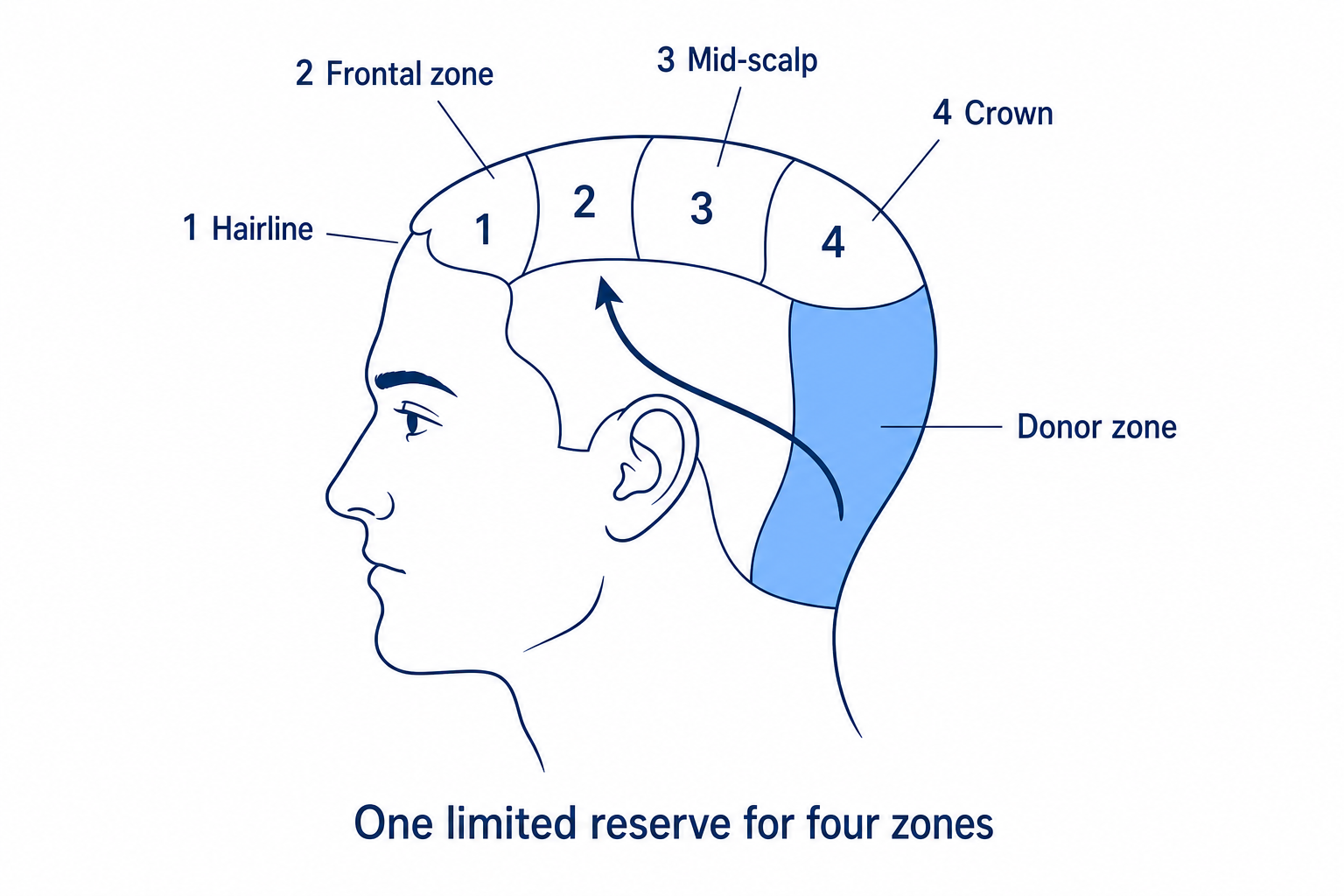
| Zone (number as in the illustration) | What it is responsible for visually | Priority in planning | Density required |
|---|---|---|---|
| 1 Hairline | Frame of the face, sets the overall impression | highest | high, fine single hairs at the front |
| 2 Frontal zone | Area directly behind the line | high | medium to high |
| 3 Mid-scalp | Transition, visually carried by the front | medium | medium, often deliberately reduced |
| 4 Crown | round area, visually more forgiving, but demanding in direction of growth | last | reduced or deliberately left out |
The crown is the most expensive square centimetre of the scalp: it runs in a circle, the direction of growth is demanding, and it visually swallows a great deal of density. The hairline, by contrast, pays off most per graft, because it forms the frame of the face. More on distribution can be found in the article on possible graft numbers.
And here is the sentence that puts the whole scale in context: the Norwood stage describes only the bald area. Whether and how well it can be covered is decided by the quality of the remaining hair, that is, donor density, hair calibre, hair texture, the contrast between hair and skin colour and head size. A Norwood 4 with thick, wavy hair is easier to plan for than a Norwood 3 with fine, straight hair.
The three biggest misconceptions about the Norwood scale
Three false assumptions about the Norwood scale come up again and again in consultations, and all three lead to poor decisions. Here they are set against the documented facts.
Misconception 1: “At some point it stops by itself.”
Male pattern hair loss runs in bursts and plateaus, and a plateau is not an end. In the Japanese cohort study of 5,372 men the median age kept rising with every stage, from 26 years at stage I to 52 years at stage VII. Whether and where a course comes to a halt cannot be predicted.
Misconception 2: “A hair transplant reverses hair loss.”
It covers area with DHT-resistant hair of your own from the donor zone. The hormonal process in the remaining hair carries on, which is why no procedure moves anyone back down the Norwood scale. Without accompanying stabilisation the pattern keeps moving around the transplanted area.
Misconception 3: “Women get a Norwood stage.”
The scale was developed on male patterns and does not fit women. A woman who looks for her own Norwood stage underestimates her situation and overlooks other, treatable causes. The Ludwig and Sinclair scales are the ones that apply.
Limits of the Norwood scale: what it cannot do
The Norwood scale is the standard, but it has known weaknesses. It captures only the pattern, not hair density, not hair calibre, not the degree of miniaturisation and not diffuse hair loss. And it depends on the observer.
The sharpest criticism has actually been measured: in the study in the International Journal of Trichology (2009) the agreement of 7 dermatologists and 16 trainee doctors on 43 photographs came to an ICC of only 0.63 to 0.68, and in a second round with 8 assessors and 56 photographs repeatability after 3 months was poor as well, with no difference between consultants and trainees.
Second weakness: the cohorts of 1951 and 1975 consisted mainly of Western men. The review in the Indian Journal of Dermatology, Venereology and Leprology shows different distributions by ethnicity, for instance type II to III as the most common classification in Indian studies, type IV in Chinese ones and type III in Korean ones.
The BASP classification (Lee WS, Ro BI, Hong SP et al., Journal of the American Academy of Dermatology 2007) is regarded as the modern alternative. It describes four basic types of hairline shape (L, M, C, U) plus two specific thinning types (F for frontal, V for vertex), works for men and women alike and represents Asian hair patterns better.
The message from all this is not to discard the scale, but to use it properly. What a stage means for you only emerges together with hair density, hair calibre, degree of miniaturisation, donor density and the speed of the course.
Ludwig scale: the classification for women
The Norwood scale is a male scale and does not fit women. Female pattern hair loss almost never begins with receding temples. It starts as diffuse thinning around the parting, with the front hairline usually preserved. The Ludwig scale with its three grades covers this (Ludwig E., British Journal of Dermatology 1977, clinical observation of 468 women).
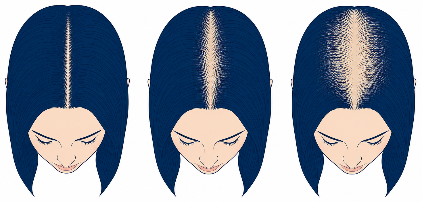
Ludwig I means slight but perceptible thinning in the front part of the crown area, with a minimally widened parting and the front hairline preserved. At Ludwig II the thinning is clearly visible, the scalp starts to show through around the parting and volume decreases noticeably.
Ludwig III describes pronounced, widespread hair loss on top of the head, with the front hairline mostly preserved here too. Precisely this preserved fringe of hair is the clear difference from the male pattern. According to the European S3 guideline (Kanti et al. 2018), 42 to 50 per cent of women are affected by pattern hair loss over the course of their lives.
Alongside Ludwig, both practice and research use the Sinclair scale, a five-point photographic system devised by Rodney Sinclair that maps the width of the central parting more finely than three grades can. A modified version achieved good to excellent inter-rater agreement (ICC 0.87) in a validation study in the Journal of the American Academy of Dermatology, considerably higher than for the Norwood scale.
In addition, E. A. Olsen described the “Christmas tree pattern”: a triangular zone of thinning that tapers towards the front, with a widened central parting. According to a review of female hair loss, this frontal emphasis is the most common female pattern, followed by central thinning similar to the male pattern.
| Scale | For whom | Levels | What it captures well | What it does not capture |
|---|---|---|---|---|
| Hamilton-Norwood | Men with pattern hair loss | 7 stages plus type A variants | Recession of the hairline and loss at the crown | Density, hair calibre, miniaturisation, diffuse forms |
| Ludwig | Women with pattern hair loss | 3 grades | Thinning around the parting with the front hairline preserved | fine intermediate steps, frontal emphasis |
| Sinclair | Women | 5 grades | the width of the central parting in finer steps | the cause of the hair loss |
| BASP | Men and women | basic type (L, M, C, U) plus specific type (F, V) | Combination of hairline shape and thinning, including Asian patterns | less widely used in everyday practice |
A woman who looks for her own Norwood stage typically underestimates her situation and overlooks other causes. Diffuse thinning can be genetic, but it can just as easily come from iron deficiency, a thyroid disorder, nutrient deficiencies, hormonal changes or telogen effluvium. These forms need a medical work-up of the cause.
A receding front hairline in a woman is a reason in its own right for a dermatological assessment, among other things because of scarring alopecias. The article on hair loss in women gives an overview of the female patterns.
Having your Norwood stage determined by a doctor
A self-assessment on the Norwood scale is enough for a first impression. For a decision you need four things that no picture comparison provides: certainty that this really is pattern hair loss, the degree of miniaturisation, the donor density actually present at the back of the head and the quality of the hair itself.
That is exactly what a medical hair analysis delivers. It determines the stage, separates pattern hair loss from diffuse hair loss, assesses the donor area and hair calibre and derives a realistic framework from all of it. At Elithair, assessing the donor zone and dividing up the recipient zones is the step that comes before any graft number.
A brief, factual word on the technique: extraction is carried out as FUE from the DHT-resistant donor zone, implantation as DHI and therefore included as standard. FUE is the extraction technique, DHI the implantation technique, and they are not alternatives to each other. If the examination shows that a procedure does not make sense right now, that is a useful result too.
Clinical perspective from the Elithair consultation
In a consultation the Norwood stage is the beginning of the conversation, not the result. It describes the bald area. Whether and how densely that area can be covered is decided by donor density, hair calibre and hair texture. These values set the upper limit, not a desired graft number.
That is why assessing the donor zone and dividing up the recipient zones comes before any figure with us. If the examination shows that a procedure does not make sense at this point, we say so.
When the Norwood scale does not fit: warning signs and differentiation
The Norwood scale describes pattern hair loss and nothing else. If hair falls out evenly across the whole head, in round patches, suddenly or together with skin complaints, then the scale is the wrong tool and a dermatological assessment is needed.
The differences in brief: a mature hairline is stable and normal. Telogen effluvium causes diffuse shedding, often after illness, a deficiency, stress or a new medication, and follows the logic of the hair cycle. Alopecia areata shows round patches, traction alopecia comes from the pull of tight hairstyles, and scarring forms go together with redness, scaling, shiny skin and the loss of the follicular openings.
Warning signs: this hair loss needs a dermatological assessment
- •very rapid hair loss within weeks
- •round, sharply defined bald patches
- •itching, burning, redness, scaling or a shiny, scarred scalp
- •a woman with a receding front hairline
- •hair loss together with fatigue, weight change or menstrual irregularities
- •hair loss that started after a new medication
Which cause lies behind it in an individual case is something no picture scale can clarify. The article on the causes of hair loss gives an overview of the possible triggers, and the practical steps that follow are covered under what to do about hair loss.
Frequently asked questions about the Norwood scale
What is the Norwood scale in one sentence?
The Norwood scale is the standard medical classification of male pattern hair loss and describes in seven stages how far the hairline has receded and how far the crown has thinned. It describes a pattern, it does not explain a cause.
How many stages does the Norwood scale have?
Seven main stages, plus the separate 3 vertex variant and the type A variants IIa to Va. Stage 1 is the youthful hairline, stage 7 leaves only a narrow rim of hair above the ears and at the nape.
Norwood or Hamilton-Norwood, what is the difference?
It is the same classification. James B. Hamilton first described the pattern systematically in 1951, and O’Tar Norwood revised it in 1975 after examining 1,000 men, adding the vertex stage and the type A variants. Norwood scale, Hamilton-Norwood scale and Norwood-Hamilton scale all mean the same thing.
Which Norwood stage am I?
Assess two zones separately and then combine them: the recession of the front hairline and the state of the crown. For that you need three photos taken in the same light, from the front, from above and in profile. The zone matrix in this article leads you to the matching stage.
Is Norwood 2 already hair loss or just a mature hairline?
On a single photo the two look the same. A mature hairline recedes once by around 1 to 1.5 centimetres during youth and then stays stable. Norwood 2 as active hair loss keeps progressing and shows thinner, shorter, lighter hairs along the edge. Only a comparison over time separates the two.
Is Norwood 2 at the age of 20 normal?
It happens and is no emergency in itself. In the Japanese cohort study of 5,372 men the median age at stage I was 26 years, so an onset at 20 is earlier than that average. It makes sense to document the progression now and have it assessed by a dermatologist.
What is the difference between Norwood 3 and Norwood 3 vertex?
At Norwood 3 the loss affects the front hairline with deep, wedge-shaped temple angles. At Norwood 3 vertex the crown thins as well, while a strip of hair still stands between the two zones. That is two areas to address instead of one, which changes the area calculation considerably.
How many grafts do you need for Norwood 3 or Norwood 4?
As a rough guide: at Norwood 3 around 1,500 to 3,000 grafts for the front zone, at Norwood 4 around 3,000 to 4,000 grafts for both zones. These are not promises. The donor area is limited, hair calibre shifts the requirement considerably, and only the medical examination is binding.
Why does one person need more grafts than another at the same stage?
Because the stage describes only the bald area. Thick, wavy hair visually covers considerably more per graft than fine, straight hair, and donor density, head size and the contrast between hair and skin colour change the calculation as well. A Norwood 4 with thick hair can be easier to plan for than a Norwood 3 with fine hair.
From which Norwood stage is a hair transplant worthwhile?
It usually becomes medically worth discussing from Norwood 3 onwards, when the loss is stable and the donor area is comfortably sufficient. At stages 1 and 2 a procedure is generally premature. From stage 5 planning becomes prioritising, and at stage 7 restoring the whole area with your own hair is usually not achievable.
Does a hair transplant stop progression on the Norwood scale?
No. Transplanted hairs come from the DHT-resistant donor zone and are generally retained, but the surrounding native hair keeps following its genetic programme. A transplant covers area, it does not change the hormonal process. That is why accompanying stabilisation is part of the planning.
What is the island effect?
It is how surgeons describe, in patient consultations, the result of a transplant into an actively progressing area: the transplanted hairs stay, the native hair around them keeps thinning, and an isolated island of hair with a bald edge appears. It is an observation from practice, not a syndrome classified in studies.
Can you go back from Norwood 3 to Norwood 2?
Not without treatment. The literature describes pattern hair loss as a progressive process that does not reverse on its own. After a transplant a zone can look denser again, but that is coverage with transplanted hair and not a biological reversal.
How quickly do you progress from one stage to the next?
In the Japanese cohort study of 5,372 men (Tokyo Memorial Clinic) the median time from one stage to the next was 4.5 years. The data come from a single clinic and a single ethnic group, and an aggressive course can be considerably faster.
Does minoxidil work better at the crown than at the hairline?
Looked at properly: the US approval for topical 5 per cent minoxidil rests on studies at the crown, and the labelling states that efficacy at the frontal hairline is not established. Newer studies such as Kanti et al. 2016 do, however, show an increase in non-vellus hairs frontotemporally as well over 104 weeks. The evidence is thinner at the front, not absent.
What is Norwood type A?
In the type A variants IIa to Va the hairline moves back evenly as a whole, without the typical wedge-shaped M form. No isolated bald spot appears at the crown; in the higher grades the front does reach the mid-scalp area, but as part of the same front. Consistent secondary sources give roughly 3 to 4 per cent of cases.
Does the Norwood scale apply to women too?
No. Female pattern hair loss almost never begins with receding temples. It starts as diffuse thinning around the parting, with the front hairline usually preserved. The Ludwig scale with its three grades covers this (Ludwig, British Journal of Dermatology 1977, 468 women).
What is the difference between the Ludwig scale and the Sinclair scale?
The Ludwig scale distinguishes three grades of thinning around the parting. The Sinclair scale works with five photographic grades and maps early changes in the width of the parting more finely. A modified Sinclair version achieved good to excellent inter-rater agreement (ICC 0.87) in a validation study in the Journal of the American Academy of Dermatology.
Is the Norwood scale a diagnosis?
No. It is a description of the visible pattern and a tool for communication between doctor, patient and study. It says nothing about cause, pace, hair density, hair calibre or degree of miniaturisation. The diagnosis comes from a medical examination.
Sources
- Norwood OT (1975): Male pattern baldness: classification and incidence. Southern Medical Journal 68(11):1359-1365 (classification based on 1,000 men examined).
- Hamilton JB (1951): Patterned loss of hair in man: types and incidence. Annals of the New York Academy of Sciences 53:708-728, PMID 14819896. pubmed.ncbi.nlm.nih.gov
- Reliability of Hamilton-Norwood Classification (2009): International Journal of Trichology (inter-rater agreement ICC 0.63 to 0.68). pmc.ncbi.nlm.nih.gov
- Age-related progression of androgenetic alopecia: Statistical analysis of 5372 Japanese men (Tokyo Memorial Clinic). pmc.ncbi.nlm.nih.gov
- Kanti V, Messenger A, Dobos G, et al. (2018): Evidence-based (S3) guideline for the treatment of androgenetic alopecia in women and in men. JEADV 32(1):11-22, PMID 29178529. pubmed.ncbi.nlm.nih.gov
- Kanti V, et al. (2016): Effect of minoxidil topical foam on frontotemporal and vertex androgenetic alopecia in men, 104-week open-label trial. JEADV, DOI 10.1111/jdv.13324. onlinelibrary.wiley.com
- British Association of Dermatologists: Patient Information Leaflet “Hair loss male pattern (androgenetic alopecia)”. bad.org.uk
- Ludwig E (1977): Classification of the types of androgenetic alopecia (common baldness) occurring in the female sex. British Journal of Dermatology 97(3):247-254 (clinical observation of 468 women).
- Lee WS, Ro BI, Hong SP, et al. (2007): A new classification of pattern hair loss that is universal for men and women: basic and specific (BASP) classification. Journal of the American Academy of Dermatology 57:37-46. pubmed.ncbi.nlm.nih.gov
- Modified Sinclair scale, validation (inter-rater agreement ICC 0.87): Journal of the American Academy of Dermatology, PubMed 34111498. pubmed.ncbi.nlm.nih.gov
- Androgenetic alopecia: An update (review of prevalence and ethnic differences). Indian Journal of Dermatology, Venereology and Leprology. ijdvl.com
- Female Pattern Hair Loss (review, Olsen pattern and female presentations). pmc.ncbi.nlm.nih.gov
- FDA Drug Label, minoxidil topical solution 5 per cent (crown indication). accessdata.fda.gov
Last reviewed 2026. This article provides general information and does not replace medical advice, diagnosis or treatment. Which stage applies in your case, and what follows from it, is something an individual medical examination will clarify.

Dr. Imad Moustafa
Hair transplant specialist